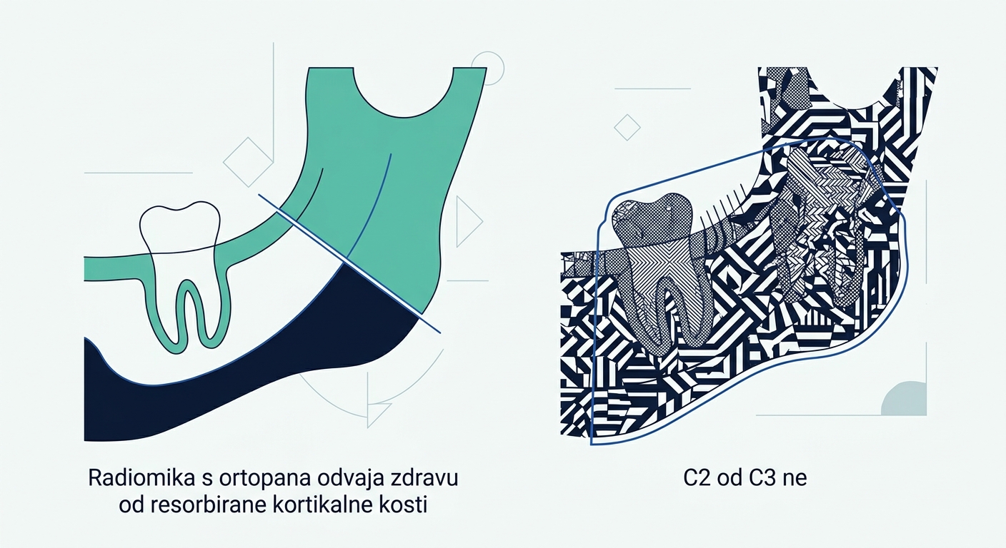
Panoramic radiomics separates intact from resorbed cortical bone, but not C2 from C3
Across 159 panoramic radiographs, radiomic features reliably separated intact cortex from resorbed cortex, but after correction for multiple testing none distinguished moderate (C2) from severe resorption (C3).
Bone texture is measurable, but not down to the final grade
The mandibular cortical index (MCI) is a simple scale: on a panoramic radiograph the appearance of the lower border of the mandible is assessed and classified as C1 (intact cortex), C2 (moderately resorbed) or C3 (severely resorbed). The assessment is visual, so it varies between observers. Uzun's team asked whether the same information could be obtained by measurement instead of by looking.
A total of 159 panoramic radiographs were analysed retrospectively, 53 in each of the three categories. The right mandibular cortex was segmented along standardised anatomical boundaries, and features from five classes — shape, first order, and the textural GLCM, GLRLM and GLSZM — were extracted from it with the PyRadiomics tool in 3D Slicer. Only features with inter-observer reliability above ICC 0.75 entered the analysis.
First-order features separated the groups consistently: energy (p = 0.006), entropy (p = 0.001), interquartile range (p = 0.002) and variance (p = 0.001) distinguished the intact cortex from the resorbed ones. The textural features behaved similarly — correlation from GLCM (p < 0.001), grey level variance from GLRLM (p = 0.001) and zone entropy from GLSZM (p = 0.002).
Then comes the sobering part. After Benjamini-Hochberg correction for multiple testing, no feature remained significantly different between C2 and C3. Measurement therefore reliably recognises that the cortex has begun to disappear, but not how far the process has gone.
Limitations
The study is retrospective and measures only the image: there is no comparison with densitometry or with clinical outcome, so it allows no conclusion about who should be referred for work-up because of bone loss. The sample of 159 radiographs is artificially balanced into three equal groups.
For your practice
- MCI remains an assessment by eye; a finding of markedly porous cortical bone on a panoramic radiograph is worth recording in the chart, not merely noticing.
- Do not spend time on finely distinguishing C2 from C3 — measurement did not manage it either once statistical correction was applied.
- If you are considering software for panoramic image analysis, ask for inter-observer reliability data; in this study only features above ICC 0.75 were eligible.