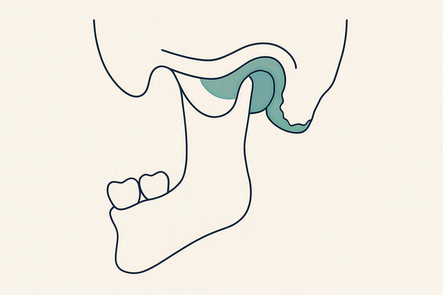
Disc displacement without reduction goes with more severe TMJ bone changes
A cross-sectional study of 198 temporomandibular joints in 99 patients showed that erosions, osteophytes and generalised sclerosis are considerably more frequent in complete disc displacement without reduction (p < 0.001).
Bone findings and disc position go together
Radiological assessment of the temporomandibular joint is split between two methods: CBCT depicts bone precisely, while magnetic resonance imaging remains the gold standard for soft tissues and the position of the articular disc. How far the two sets of findings actually coincide had not been clarified.
The team led by Matea Prenc published in the journal Acta Stomatologica Croatica a cross-sectional study of 198 temporomandibular joints in 99 patients with a clinical diagnosis of osteoarthritis. Every patient was imaged with both methods, so the findings can be compared directly. Disc position on MRI was sorted into four categories: normal, partial displacement with reduction, complete displacement with reduction, and complete displacement without reduction. CBCT recorded subchondral cysts, erosions, generalised sclerosis, osteophytes and loose joint bodies, and the degree of osteoarthritis was determined according to the Ahmad and Schiffman criteria. Differences were tested with Fisher's exact test.
Joints with complete displacement without reduction stood apart from the rest on almost every bone finding. Erosions, generalised sclerosis and osteophytes were significantly more frequent there (all p < 0.001), and the same holds for subchondral cysts (p = 0.004). They most often coincided with grade two osteoarthritis (26.8%), so the relationship between the degree of progression and the type of displacement also turned out to be statistically significant (p < 0.001). The only finding that did not fit that pattern was loose joint bodies: their presence did not depend on disc position (p = 0.191).
Limitations
This is a cross-sectional study in a single group of patients, so the finding speaks of an association, not of a causal sequence. The data do not show whether disc displacement precedes bone breakdown or follows it.
For your practice
- Treat a finding of displacement without reduction on MRI as an indicator of an advanced stage and expect more pronounced bone remodelling, even when the bone findings on CBCT are only moderate.
- One modality is not enough for planning treatment of TMJ osteoarthritis; CBCT and MRI provide information that cannot substitute for one another.
- A normal CBCT does not rule out internal derangement of the joint, and loose joint bodies are not a usable indicator of disc position.